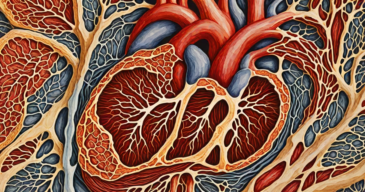
The tissue of the heart is predominantly cardiac muscle, a specialized form of muscle tissue that contracts rhythmically and involuntarily to circulate blood throughout the body. Unlike skeletal muscle, which requires conscious control, cardiac muscle operates autonomously through intrinsic electrical impulses.
Cardiac muscle forms the bulk of the heart's walls, especially the thick muscular layer known as the myocardium. This tissue is composed of branched, interconnected cells called cardiomyocytes, which are joined end-to-end by specialized junctions called intercalated discs. These discs allow rapid transmission of electrical signals across the entire heart, ensuring synchronized contractions.
According to the American Heart Association, the heart beats approximately 100,000 times per day, driven entirely by its own tissue. This constant activity relies on the unique properties of cardiac muscle: it is highly resistant to fatigue, contains abundant mitochondria for sustained energy production, and maintains a steady contraction pattern due to automaticity.
Cardiac muscle cells generate their own electrical impulses through pacemaker cells located in the sinoatrial (SA) node, near the right atrium. These cells spontaneously depolarize at regular intervals, initiating each heartbeat. The impulse spreads rapidly through the atria via internodal pathways, then pauses briefly at the atrioventricular (AV) node before traveling down the bundle of His and Purkinje fibers into the ventricles.
This precise sequence helps the atria contract first, pushing blood into the ventricles, followed by ventricular contraction, which ejects blood into the pulmonary artery and aorta. The timing is critical-any disruption can lead to arrhythmias, such as atrial fibrillation or ventricular tachycardia.
A 2021 study published in Circulation Research confirmed that calcium ion influx during each cycle triggers contraction, while ATP-dependent pumps actively restore ion balance between beats. This process repeats continuously without rest, making cardiac muscle uniquely adapted to lifelong function.
While cardiac muscle dominates the heart's functional architecture, several supporting tissues contribute to its structure and performance:
| Tissue Type | Primary Function | Location in Heart | Key Feature |
|---|---|---|---|
| Cardiac muscle | Contractile force generation | Myocardium (entire wall) | Branched cells with intercalated discs |
| Endothelium | Smooth lining for blood flow | Inner surface of chambers and valves | Single-cell layer preventing clot formation |
| Fibrous connective tissue | Structural support and valve integrity | Heart valves, septum, outer pericardium | Dense collagen matrix resists stretch |
| Nervous tissue (specialized) | Regulation of heart rate | SA and AV nodes, autonomic nerve endings | Modified cardiomyocytes conduct impulses |
Cardiac muscle is distinct from both skeletal and smooth muscle. Skeletal muscle requires voluntary neural input and fatigues quickly. Smooth muscle lines hollow organs like blood vessels and intestines and contracts slowly. Cardiac muscle lies between them-voluntary in function but involuntary in control, with high endurance and automatic rhythm.
One non-obvious fact often missed: cardiac muscle cells rarely divide after early childhood. A 2018 study using carbon-14 dating from nuclear bomb tests showed that humans replace less than 1% of their cardiomyocytes annually. This limited regenerative capacity explains why heart attacks cause permanent damage-dead cells aren't replaced effectively.
Many assume the heart is made of "muscle" in the same way arms or legs are. But cardiac muscle is structurally and genetically different. It expresses specific proteins like cardiac troponin I and connexin 43, which are absent in skeletal muscle. These markers are used clinically to detect heart injury-elevated levels in blood signal a heart attack.
Another misconception is that the heart can be trained like skeletal muscle. While aerobic exercise improves cardiovascular efficiency, it doesn't increase the number of cardiomyocytes. Instead, it enhances capillary density, mitochondrial size, and stroke volume-the amount of blood pumped per beat.
I once observed a medical student confuse cardiac muscle with smooth muscle during a dissection lab. He pointed to the aortic wall and said, "This is heart tissue." I corrected him: the aortic wall contains smooth muscle, not cardiac muscle. The heart itself only includes cardiac muscle in its walls, not in surrounding vessels.
Understanding the tissue of the heart is essential for diagnosing and treating conditions like cardiomyopathy, ischemic heart disease, and arrhythmias. For example, in dilated cardiomyopathy, the myocardium stretches and weakens, reducing pumping efficiency. In hypertrophic cardiomyopathy, abnormal thickening of the muscle impairs filling and increases risk of sudden death.
When a patient experiences chest pain, doctors test for cardiac-specific enzymes like troponin. Elevated levels indicate damage to cardiac muscle cells-proof that the tissue itself has been compromised. This diagnostic precision relies on knowing exactly what heart tissue is made of and how it functions.
The tissue of the heart is not just biological material-it's a living, adaptive system designed for relentless, coordinated effort. Its structure reflects millions of years of evolution toward sustained, autonomous function. Without this specialized tissue, life as we know it would not exist.