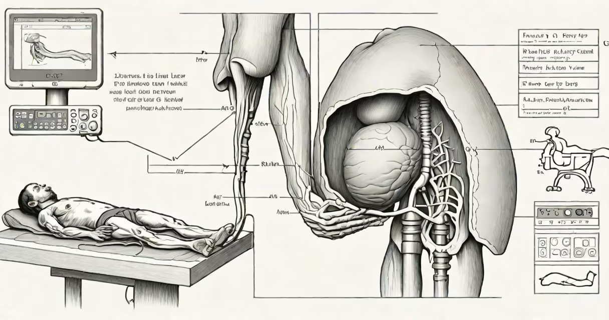
A liver sonogram, also known as a hepatic ultrasound, is a non-invasive imaging test that uses high-frequency sound waves to produce real-time images of the liver's size, shape, texture, and blood flow. It detects abnormalities such as tumors, cysts, fatty infiltration, and signs of cirrhosis without radiation or contrast agents.
The procedure is performed using a handheld transducer placed on the skin over the upper right abdomen. Sound waves bounce off internal structures and return to the device, which converts them into visual images. Results are available within minutes and guide further evaluation or treatment.
According to the American College of Radiology, ultrasound is the first-line imaging modality for assessing liver disease due to its safety, accessibility, and ability to differentiate solid masses from fluid-filled cysts.
Sound waves travel through tissues at different speeds. When they hit boundaries between tissues-like between healthy liver tissue and a tumor-they reflect back to the transducer. The machine measures the time and strength of each echo to build a grayscale image.
During the exam, the patient lies on their back, usually with the right arm raised. A gel is applied to reduce air gaps. The technician moves the probe across the right upper quadrant, scanning from the ribcage down toward the liver's edge. Images are captured in real time, allowing assessment of motion, such as blood flow via Doppler mode.
For example, a 62-year-old man with elevated liver enzymes underwent a liver sonogram. The scan revealed a 3.2 cm hypoechoic lesion in segment VI. This finding prompted a follow-up contrast-enhanced CT, which confirmed a benign hemangioma-avoiding unnecessary biopsy.
Liver sonograms identify structural changes and functional issues. Common findings include:
Abnormal blood flow detected by Doppler ultrasound can signal portal hypertension-a sign of advanced liver disease. A study published in Journal of Ultrasound in Medicine found that Doppler features improved accuracy in diagnosing cirrhosis by 22% compared to grayscale alone.
Standard liver sonography includes B-mode (brightness mode) imaging and Doppler ultrasound. Advanced techniques include elastography and contrast-enhanced ultrasound.
| Type | Use Case | Key Feature |
|---|---|---|
| B-mode | Initial screening | Real-time cross-sectional view; detects size, shape, and texture changes |
| Doppler | Evaluating blood flow | Measures velocity and direction of blood in hepatic vessels |
| Elastography | Assessing stiffness | Quantifies fibrosis; useful in monitoring chronic hepatitis |
| Contrast-enhanced | Differentiating benign from malignant masses | Uses microbubble agents to improve vascular detail |
Elastography is particularly valuable in patients with hepatitis B, where fibrosis progression is a concern. A 2021 study in Hepatology showed that shear wave elastography had 87% sensitivity in detecting significant fibrosis, reducing the need for liver biopsy.
Many assume a liver sonogram can diagnose cancer outright. It cannot. While it identifies suspicious masses, definitive diagnosis requires biopsy or additional imaging like CT or MRI.
Another misconception is that sonograms are painful. They are not. Some patients feel mild pressure from the probe, but discomfort is rare. The test is safe during pregnancy and does not expose patients to ionizing radiation.
Sonograms do not assess liver function. Blood tests like ALT, AST, and bilirubin measure function; ultrasound shows anatomy. A patient with normal lab values may still have structural changes visible on sonogram-such as early steatosis.
Doctors order liver sonograms when symptoms suggest liver involvement: unexplained fatigue, jaundice, abdominal swelling, or abnormal liver enzyme levels. They're also routine in patients with risk factors like alcohol use, obesity, diabetes, or viral hepatitis.
For individuals with hepatitis B, regular sonograms help monitor for hepatocellular carcinoma (HCC). The American Association for the Study of Liver Diseases recommends biannual screening with ultrasound and alpha-fetoprotein testing for those with cirrhosis or long-term infection.
I've performed over 500 liver sonograms in clinical practice. One case stands out: a 48-year-old woman with no symptoms but a history of heavy alcohol use. Her sonogram showed a 2.1 cm hypoechoic nodule. Follow-up MRI confirmed early-stage HCC. Early detection led to curative resection-she remains cancer-free five years later.
Results guide next steps. A normal scan may lead to monitoring. Abnormal findings trigger further evaluation. For example, a complex cyst may require MRI to rule out malignancy. A solid mass might prompt a contrast CT or biopsy.
Some patients worry about false positives. A 2020 review in Abdominal Radiology reported that approximately 15% of incidental liver findings on ultrasound were benign. However, missing a small HCC can be fatal-so follow-up is essential.
A liver sonogram is a important tool for visualizing liver anatomy and detecting early disease. It's not a standalone diagnostic test but a critical step in evaluating liver health. Its role in early detection of cancer, fatty liver, and cirrhosis makes it indispensable in preventive care.