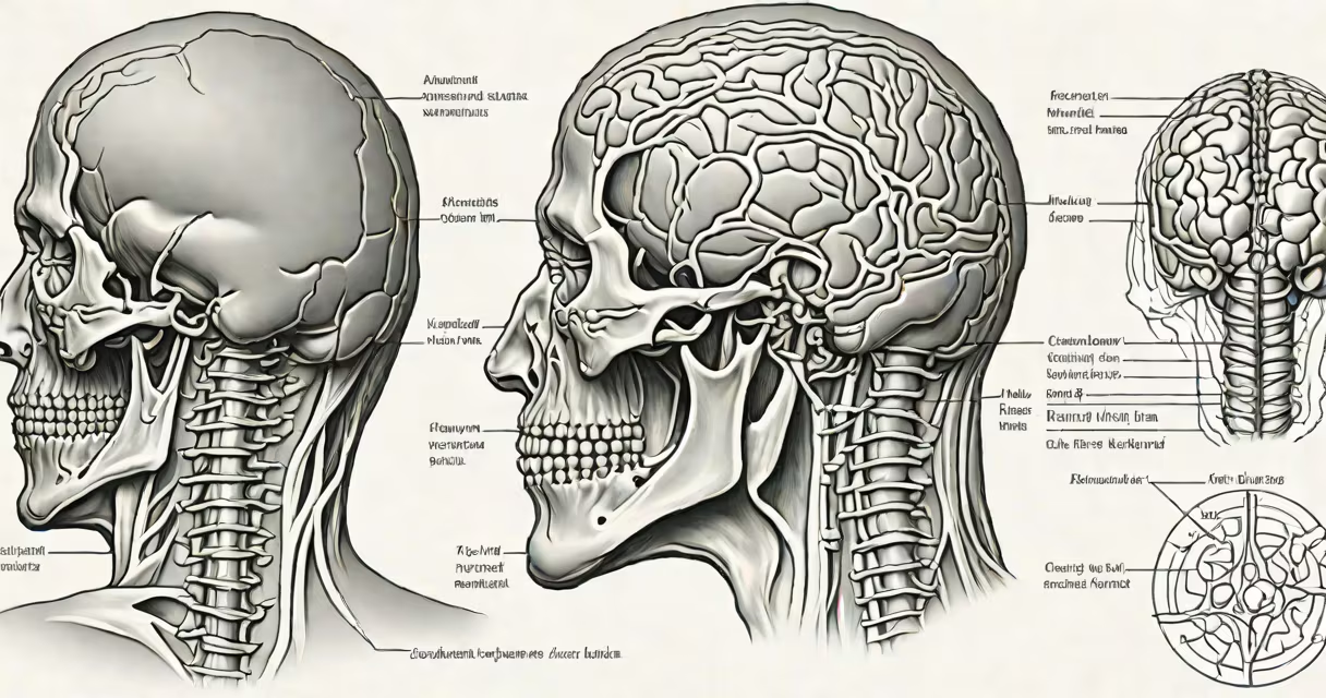
A herniated disc in back and neck happens when the gel-like center of a spinal disc ruptures through its tough outer layer, compressing nearby nerves. This condition often causes sharp pain, tingling, or muscle weakness along the path of the affected nerve. It most commonly affects the lower back (lumbar region) and upper neck (cervical region).
Spinal discs act as shock absorbers between vertebrae. Each disc consists of a fibrous outer ring called the annulus fibrosus and a soft, jelly-like nucleus pulposus at its core. When trauma, repetitive strain, or age-related degeneration weakens the outer layer, the nucleus can push outward. If it breaks through, it becomes a herniated disc.
According to the American Academy of Orthopaedic Surgeons (AAOS), approximately 1% of people develop a symptomatic lumbar disc herniation each year. In the cervical spine, the rate is lower but still significant, especially among individuals aged 30-50 who perform frequent overhead lifting or have prolonged poor posture.
Herniations typically occur due to cumulative stress rather than sudden injury. Repetitive bending, twisting, or heavy lifting increases pressure on the disc's posterior side. Over time, this leads to micro-tears in the annulus, allowing the nucleus to protrude. Once the material contacts a nerve root, symptoms emerge.
The lumbar spine (lower back) handles about 70% of the body's weight during standing and walking. The L4-L5 and L5-S1 discs are most vulnerable. In the cervical spine (neck), the C5-C6 and C6-C7 levels are common sites. A herniation at C6-C7 may compress the C7 nerve root, leading to pain radiating down the arm into the middle finger.
For example, a warehouse worker who lifts boxes daily may develop a lumbar herniation after years of improper form. MRI imaging reveals a 6 mm posterior disc protrusion at L5-S1, impinging on the S1 nerve root. The patient reports burning pain from the lower back down the back of the left leg, worsening when sitting or coughing.
Disc herniations are categorized by location and severity:
| Type | Description | Common Location | Typical Symptoms |
|---|---|---|---|
| Protrusion | Inner material pushes against the outer wall but remains contained | Lumbar and cervical spine | Mild pain, stiffness, occasional numbness |
| Prolapse | Disc material extends beyond the normal boundary but is still connected | Cervical spine (C5-C6) | Neck pain, shoulder weakness, tingling in index finger |
| Rupture | Complete breach of the annulus; nucleus escapes freely | Lumbar spine (L4-L5) | Severe sciatica, foot drop, loss of reflexes |
Not all herniations cause symptoms. A study published in Spine Journal found that 30% of asymptomatic adults aged 30-50 had disc herniations visible on MRI scans. This shows that structural changes do not always equate to clinical problems.
Many assume a herniated disc means permanent damage or requires surgery. That is incorrect. Most cases resolve within six weeks with conservative care. The National Institute of Neurological Disorders and Stroke (NINDS) states that 90% of patients improve without surgical intervention.
Another misunderstanding is equating disc herniation with sciatica. Sciatica refers specifically to pain along the sciatic nerve, which runs from the lower back through the buttocks and down the leg. While a lumbar herniation can cause sciatica, not all sciatica stems from disc issues-piriformis syndrome or spinal stenosis can mimic the same pattern.
Also, herniations are not always caused by physical trauma. Degenerative disc disease, which begins in the late 20s, gradually reduces disc hydration and elasticity. By age 40, nearly half of adults show signs of disc degeneration on imaging, increasing the risk of herniation even without injury.
Diagnosis begins with a neurological exam assessing strength, sensation, and reflexes. Imaging confirms the presence and location of the herniation. An MRI is the gold standard-it visualizes soft tissue, including nerve roots and disc material. CT scans with myelography are used when MRI is contraindicated (e.g., pacemaker users).
Treatment follows a stepwise approach. First-line therapy includes NSAIDs, physical therapy focused on core and neck stabilization, and activity modification. A 2022 Cochrane review concluded that supervised exercise programs improved function and reduced pain in 70% of participants after eight weeks.
If symptoms persist beyond six weeks, corticosteroid injections may be considered. Epidural steroid injections reduce inflammation around the nerve root and provide temporary relief. A randomized trial in JAMA Neurology showed a 50% reduction in leg pain at three months compared to placebo.
Surgery is reserved for severe cases. Microdiscectomy removes the portion of the disc pressing on the nerve. Success rates exceed 90% for properly selected patients. However, risks include infection, dural tear, and recurrent herniation (about 5-10% within five years).
I once treated a 42-year-old office worker with a C6-C7 herniation causing numbness in the thumb and index finger. After four weeks of physical therapy focusing on cervical extension and scapular control, symptoms resolved. MRI follow-up showed no progression. This case illustrates that many patients recover without invasive procedures.
Seek medical evaluation if pain radiates below the waist or down the arm, especially with weakness or loss of bladder/bowel control. These indicate cauda equina syndrome-a rare but serious condition requiring emergency surgery.
Prevention focuses on posture, movement habits, and load management. Avoid prolonged sitting with the head forward. Use ergonomic chairs and take breaks every 30 minutes. Strengthen the deep neck flexors and transverse abdominis muscles through targeted exercises.
Understanding herniated disc in back and neck as a mechanical issue-not a catastrophic failure-empowers informed decisions. With proper care, most people return to normal activities within months.