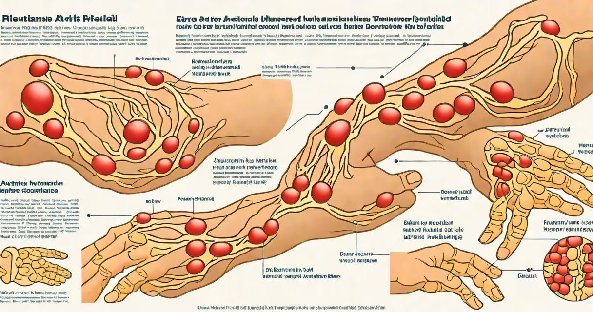
Extra articular manifestations of rheumatoid arthritis are clinical features that occur outside the joints in people with rheumatoid arthritis, indicating systemic disease activity. They include skin nodules, lung fibrosis, vasculitis, and neurological or ocular involvement. These manifestations are not caused by joint damage but reflect immune system dysfunction affecting multiple organs.
Rheumatoid arthritis is an autoimmune disorder where the immune system attacks synovial tissue in joints. While joint inflammation defines the condition, extra articular manifestations arise when autoimmunity extends beyond the musculoskeletal system. These events occur in approximately 20-30% of patients with long-standing or severe disease, according to the American College of Rheumatology.
These manifestations are not merely side effects-they are markers of disease severity and predictors of poor outcomes. Patients with extra articular features often have higher levels of rheumatoid factor and anti-CCP antibodies, suggesting more aggressive immune activation. Early recognition improves management and reduces mortality risk.
Extra articular manifestations stem from immune complex deposition, chronic inflammation, and vascular injury. Autoantibodies such as rheumatoid factor and anti-citrullinated protein antibodies (ACPAs) form immune complexes that deposit in tissues, triggering inflammation and tissue damage. This process can affect blood vessels, lungs, skin, and nerves.
For example, rheumatoid nodules form when macrophages and fibroblasts accumulate in subcutaneous tissue, often near pressure points like elbows or heels. These nodules are not painful but indicate persistent immune activation. A study published in *Arthritis & Rheumatology* found that nodules were associated with higher disease activity scores and increased risk of pulmonary complications.
Another mechanism involves endothelial cell damage. In vasculitis, immune complexes attack small blood vessels, leading to ischemia and tissue necrosis. This can result in digital ulcers, skin infarcts, or peripheral neuropathy. The presence of vasculitis increases morbidity and requires aggressive immunosuppression.
The most frequently observed extra articular manifestations include cutaneous nodules, pulmonary involvement, and vasculitis. Less common but clinically significant are ocular, neurological, and cardiac manifestations.
| Type | Prevalence | Key Features | Associated Risk Factors |
|---|---|---|---|
| Skin nodules | 10-20% | Subcutaneous, firm, often over bony prominences; may ulcerate | Rheumatoid factor positive, high anti-CCP titers, smoking history |
| Pulmonary fibrosis | 5-10% | Progressive dyspnea, interstitial lung disease on HRCT | Long disease duration, male sex, smoking |
| Vasculitis | 1-2% | Digital ulcers, livedo reticularis, mononeuritis multiplex | High disease activity, seropositive status |
| Ocular dryness (Sjögren's overlap) | 15-20% | Recurrent keratoconjunctivitis sicca, corneal ulcers | Anti-Ro/SSA positivity |
One non-obvious fact: rheumatoid nodules are not always benign. While most are asymptomatic, up to 10% can undergo malignant transformation into sarcomas, particularly in patients with long-standing disease. The risk is low but warrants biopsy if a nodule changes in size or texture.
A patient with longstanding rheumatoid arthritis presented with progressive shortness of breath and a dry cough. High-resolution CT scan revealed bilateral ground-glass opacities and traction bronchiectasis. Lung biopsy confirmed non-specific interstitial pneumonia with fibrosis. This case illustrates how pulmonary involvement can mimic infection or sarcoidosis, delaying diagnosis unless clinicians consider extra articular disease.
These manifestations typically emerge in patients with persistent, uncontrolled disease. They are more common in men, smokers, and those with high-titer autoantibodies. Smoking is a major modifiable risk factor-studies show it doubles the likelihood of developing nodules and lung disease.
They appear because rheumatoid arthritis is not solely a joint disease. It is a systemic inflammatory condition. The immune system's misdirected attack affects connective tissues throughout the body. For example, the same cytokines driving joint destruction-TNF-alpha, IL-6, and IFN-gamma-also promote fibrosis in the lungs and endothelial damage in blood vessels.
Patients with extra articular features often have higher rates of cardiovascular events. A 2021 cohort study in *The Lancet Rheumatology* found that RA patients with vasculitis had a 3.2-fold increased risk of myocardial infarction compared to those without systemic involvement.
Many assume extra articular manifestations are rare or only seen in advanced cases. In reality, they can appear early. A 2019 study in *Annals of the Rheumatic Diseases* reported that 15% of newly diagnosed patients had at least one extra articular feature within the first year.
Another misconception is that all skin changes in RA are nodules. Some patients develop rashes due to medications (e.g., methotrexate-induced erythema multiforme), while others have overlapping conditions like psoriasis or lupus. Distinguishing between drug reactions, secondary autoimmune diseases, and true RA-related skin manifestations is critical.
True rheumatoid nodules are typically painless, firm, and located over pressure points. They do not fluctuate with joint swelling. In contrast, drug-induced rashes often appear suddenly, are pruritic, and resolve after stopping the medication. Misdiagnosis leads to inappropriate treatment.
I once evaluated a 58-year-old woman with a 12-year history of rheumatoid arthritis who developed a new nodule on her right elbow. Biopsy confirmed granulomatous inflammation with central necrosis-classic for rheumatoid nodules. She was not on biologics at the time. After initiating rituximab, the nodule regressed over six months. This case highlights that systemic therapy can reverse some extra articular features, not just suppress joint inflammation.
Diagnosis relies on clinical suspicion, imaging, and biopsy. A patient with unexplained weight loss, fever, or new neurological deficits should prompt screening for systemic RA. Tests include chest CT for lung disease, nerve conduction studies for neuropathy, and skin biopsy for nodules.
Management focuses on controlling systemic inflammation. Disease-modifying antirheumatic drugs (DMARDs) like methotrexate and leflunomide are first-line. For severe cases, biologic agents targeting TNF-alpha (e.g., infliximab) or B cells (e.g., rituximab) are effective. Corticosteroids may be used short-term for acute flares.
Early intervention prevents irreversible damage. For example, pulmonary fibrosis progresses rapidly once established. Treatment with tocilizumab (IL-6 inhibitor) has shown stabilization in some patients, though evidence remains limited.
Extra articular manifestations correlate with reduced quality of life and increased mortality. The presence of vasculitis or severe lung disease reduces five-year survival by nearly 20%, according to data from the UK Biobank cohort.
Regular monitoring is essential. Patients should report new symptoms such as vision changes, chest pain, or foot numbness. Annual chest imaging and ophthalmologic exams are recommended for those with known systemic disease.
Recognizing extra articular manifestations of rheumatoid arthritis is not optional-it's necessary for complete care. These features signal that the disease is active beyond the joints and demand systemic treatment. Early detection and aggressive management improve outcomes and prevent disability.