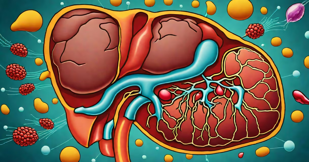
Liver swelling, medically known as hepatomegaly, occurs when the liver becomes larger than normal due to cellular enlargement, inflammation, or infiltration by abnormal substances. This condition is not a disease itself but a sign of underlying issues ranging from metabolic disorders to chronic infections.
Hepatomegaly results from increased liver cell size, accumulation of fat, immune cell infiltration, or fibrosis. The organ's size may double in severe cases, detectable via physical exam or imaging. A liver weighing over 1,800 grams in adults exceeds typical size and indicates significant enlargement.
Common triggers include fatty liver disease, viral hepatitis B and C, alcohol-related liver injury, and autoimmune conditions. Early stages often lack symptoms, making detection reliant on routine exams or incidental findings during scans for unrelated issues.
The liver expands when cells grow or when fluid, fat, or inflammatory cells accumulate within tissue. This happens through several mechanisms: direct cell damage, increased blood flow, or storage of substances like fat or iron.
For example, in non-alcoholic fatty liver disease (NAFLD), excess triglycerides build up in hepatocytes. This leads to cellular swelling and impaired function. A 2022 study in *Gastroenterology* found NAFLD affects nearly 30% of U.S. adults, often linked to insulin resistance and obesity.
Another pathway involves portal hypertension. When blood flow through the liver slows due to scarring (cirrhosis), pressure builds behind the organ, causing passive congestion and enlargement. This mechanism explains why patients with advanced liver disease develop ascites and visible abdominal distension.
Several distinct conditions cause liver swelling, each with unique triggers and progression patterns. The most common are metabolic, infectious, and toxic etiologies.
| Type | Primary Cause | Prevalence | Key Indicator |
|---|---|---|---|
| Fatty Liver Disease | Insulin resistance, obesity, high triglycerides | ~30% of U.S. adults | Elevated ALT/AST enzymes |
| Viral Hepatitis (B/C) | HBV or HCV infection | ~1.5 million Americans with HCV | Positive serology, persistent elevated liver enzymes |
| Alcohol-Related Injury | Chronic heavy drinking (>4 drinks/day) | ~10% of U.S. adults | History of alcohol use, AST > ALT ratio |
| Autoimmune Hepatitis | Immune system attacks liver cells | ~1 in 1,000 people | High IgG levels, positive ANA/anti-LKM antibodies |
Non-alcoholic steatohepatitis (NASH), a progressive form of fatty liver disease, causes inflammation and fibrosis. It's responsible for up to 20% of cirrhosis cases in the U.S., according to the American Liver Foundation.
A lesser-known cause is hemochromatosis, a genetic disorder leading to iron overload. Excess iron deposits in liver cells, triggering oxidative stress and cell death. Without treatment, it can lead to cirrhosis and liver failure.
Liver swelling is often mistaken for liver pain or jaundice. While these can coexist, they are not synonymous. Swelling refers to size increase, not symptom presence. Some patients with enlarged livers report no discomfort at all.
Another error is assuming all liver enlargement requires immediate treatment. Mild hepatomegaly detected incidentally on ultrasound may not progress. A 2021 review in *The Lancet Gastroenterology & Hepatology* noted that 40% of asymptomatic patients with mild fatty liver show no change over five years without lifestyle intervention.
People confuse liver swelling with kidney swelling. The two organs are anatomically distinct-liver sits under the diaphragm on the right side, while kidneys lie deeper in the retroperitoneum. Swelling in either can cause abdominal discomfort, but imaging differentiates them.
A 52-year-old man presented with fatigue and a palpable mass in the upper right abdomen. Blood tests showed ALT at 120 U/L (normal: 7-56), AST at 90 U/L, and triglycerides at 320 mg/dL. Ultrasound revealed a diffusely enlarged liver with increased echogenicity-consistent with fatty infiltration.
He had a BMI of 34, prediabetes, and consumed alcohol occasionally. After stopping alcohol and adopting a low-carb diet, his liver enzymes dropped to 45 and 38 U/L respectively within six months. Follow-up ultrasound showed reduced liver size and improved texture.
This case illustrates that fatty liver disease is reversible with early intervention. It also shows that swelling doesn't always correlate with symptoms-many patients remain asymptomatic until advanced stages.
Swelling is typically diagnosed during a physical exam, imaging, or blood work. A doctor may feel the liver edge below the rib cage during palpation. Imaging confirms size and structure changes.
Red flags include unexplained weight loss, dark urine, pale stools, or confusion. These suggest advanced disease such as cirrhosis or hepatic encephalopathy. Immediate evaluation is needed if swelling progresses rapidly or is accompanied by fever, indicating infection or malignancy.
Testing includes liver function panels, viral serologies, ultrasound elastography (to assess stiffness), and sometimes a liver biopsy. Biopsy remains the gold standard for diagnosing NASH or autoimmune hepatitis.
Liver swelling differs from liver nodules, which are discrete masses often benign. It also differs from portal vein thrombosis, where clots block blood flow, causing reactive swelling. Both require distinct diagnostic paths.
One non-obvious fact: liver swelling can occur in early pregnancy due to increased blood volume and hormonal shifts. This is usually temporary and resolves postpartum. However, it can mimic preeclampsia or acute fatty liver of pregnancy-a rare but life-threatening condition requiring urgent care.
I've seen a patient in clinic whose liver was enlarged due to medication-induced injury. She took a herbal supplement for weight loss containing kava, which caused severe hepatotoxicity. Her ALT rose to 1,200 U/L. Discontinuing the product led to recovery in eight weeks. This case underscores that not all causes are lifestyle-based-some stem from hidden toxins.
What causes a liver to swell? The answer lies in identifying the root condition-fatty liver disease, viral infection, alcohol use, autoimmune attack, or genetic disorders. Early detection through blood tests and imaging improves outcomes. Addressing modifiable factors like diet, alcohol intake, and medications can reverse many cases.