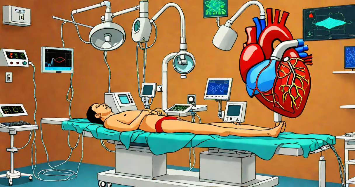
Catheter ablation for supraventricular tachycardia (SVT) is a minimally invasive procedure that destroys small areas of heart tissue responsible for causing abnormal electrical signals that trigger rapid heartbeats. It's performed under local anesthesia and sedation, with a thin tube (catheter) threaded through blood vessels to the heart.
SVT occurs when electrical impulses in the heart's upper chambers fire too quickly or follow abnormal paths. This leads to episodes of palpitations, dizziness, chest pain, or fainting. Ablation targets the specific site-such as the AV node, accessory pathways, or atrial foci-where the arrhythmia originates. By applying radiofrequency energy or cryoenergy, the procedure creates tiny scars that block faulty signals.
The American Heart Association reports that ablation for SVT has a success rate exceeding 95% for most common forms, including AV nodal reentrant tachycardia (AVNRT) and Wolff-Parkinson-White syndrome. Most patients resume normal activities within 24 to 48 hours post-procedure.
Ablation begins with mapping the heart's electrical activity using catheters placed in the femoral vein and advanced into the right atrium. Specialized electrodes record signal timing and origin. Once the abnormal pathway is located, the ablation catheter delivers controlled energy to destroy the tissue.
Radiofrequency ablation uses heat (around 60-70°C) to create precise lesions. Cryoablation uses cold (down to −40°C) to freeze tissue, which may reduce risk of damage to nearby structures like the phrenic nerve. Both methods are effective, but cryoablation is preferred in cases near sensitive areas.
During the procedure, the patient remains awake but sedated. Real-time fluoroscopy and electroanatomical mapping systems (like CARTO or EnSite) guide the physician. The entire process takes 60 to 90 minutes, depending on complexity.
Not all SVTs respond the same way to ablation. The most common types include:
| Type | Target Tissue | Success Rate | Common Symptoms |
|---|---|---|---|
| AV nodal reentrant tachycardia (AVNRT) | Slow pathway in AV node | 97-99% | Sudden onset palpitations, lightheadedness |
| Wolff-Parkinson-White (WPW) | Accessory pathway (bundle of Kent) | 95-98% | Palpitations, syncope, risk of sudden death during tachycardia |
| Orthodromic AV reentrant tachycardia (AVRT) | Anterograde conduction via AV node, retrograde via accessory pathway | 95% | Regular rapid heartbeat, often triggered by stress or caffeine |
| Atypical atrial flutter | Re-entry circuit in right atrium | 90-93% | Fluttering sensation, fatigue, shortness of breath |
Each type requires different mapping strategies. For example, WPW patients often show a delta wave on ECG during sinus rhythm, indicating an early activation of the ventricles. This sign helps confirm the diagnosis before ablation.
Ablation is recommended when medications fail to control symptoms or cause side effects. It's also advised for patients who want to avoid long-term drug therapy. The procedure is typically offered after a formal electrophysiology study confirms the mechanism of the arrhythmia.
Guidelines from the Heart Rhythm Society state that ablation should be considered early in symptomatic patients with documented SVT, especially those whose quality of life is affected. A 2021 study in Journal of the American College of Cardiology found that patients undergoing ablation had better symptom relief and reduced healthcare costs compared to those managed with drugs alone.
Procedures are performed in specialized cardiac electrophysiology labs. Centers with high-volume experience report lower complication rates. The National Cardiovascular Data Registry shows that centers performing more than 100 ablations annually have a 1.2% procedural complication rate versus 3.5% in low-volume centers.
Many assume ablation is a last resort. In reality, it's often the first-line treatment for recurrent SVT. The procedure is not experimental-it's well-established and recommended in major clinical guidelines.
Another myth is that ablation always causes permanent heart block. While AV nodal ablation can lead to complete heart block requiring a pacemaker, this is rare and only done when other options fail. Modern techniques aim to preserve AV nodal function whenever possible.
Some patients fear radiation exposure. Fluoroscopy is used, but modern systems minimize dose. A 2020 study in Circulation: Arrhythmia and Electrophysiology showed that average radiation exposure during SVT ablation is 5-8 mSv-equivalent to about 1-2 years of natural background radiation.
She experienced palpitations lasting 15-30 minutes, occurring 2-3 times weekly. Her ECG confirmed AVNRT. Medications caused fatigue and dizziness. She underwent ablation at a tertiary center.
During the procedure, the electrophysiologist mapped the slow pathway in the AV node. Radiofrequency energy was applied for 30 seconds at 45 watts. The abnormal pathway was eliminated. No recurrence occurred during 18 months of follow-up. She returned to work within 48 hours.
This case illustrates the typical outcome: rapid symptom resolution, minimal downtime, and high long-term success.
I observed an ablation for AVNRT in a teaching hospital. The team used a 3D mapping system to visualize the heart's anatomy in real time. The patient reported feeling no pain during ablation but described a brief warm sensation when energy was delivered. After the procedure, she walked out the same day with no complications.
Patients rest for 4-6 hours post-procedure. The groin site is monitored for bleeding. Most return home the same day. Avoid heavy lifting for 24 hours and swimming for five days.
Follow-up includes a 24-hour Holter monitor at one month to check for recurrence. If no symptoms or arrhythmia are detected, further testing is usually unnecessary. Long-term outcomes remain excellent-over 90% of patients remain free of SVT at five years.