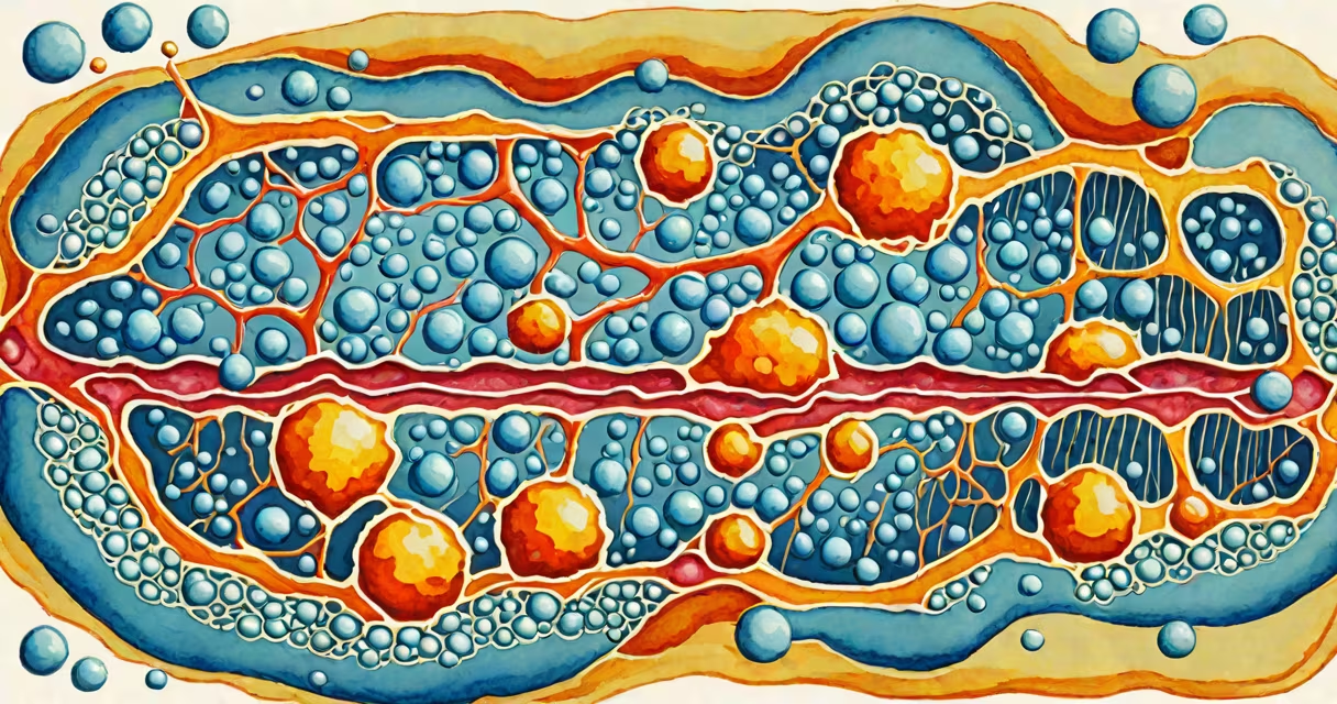
The main component of the plasma membrane is the phospholipid bilayer-a double layer of amphipathic molecules with hydrophilic heads facing outward and hydrophobic tails oriented inward. This structure creates a stable, semi-permeable barrier that separates the cell's interior from its external environment.
Each phospholipid molecule consists of a glycerol core, two fatty acid chains, and a phosphate-containing head group. In aqueous environments, these molecules spontaneously arrange into bilayers due to their amphipathic nature. The hydrophilic heads interact with water on both sides of the membrane, while the hydrophobic tails face each other in the center, shielding themselves from water.
This arrangement forms the foundation of cellular boundaries. Without it, cells could not maintain internal conditions necessary for life. The bilayer is not static-it exhibits lateral movement, allowing changing interactions with embedded proteins and lipids. This fluidity is essential for processes like membrane fusion during exocytosis and endocytosis.
The phospholipid bilayer is primarily composed of phosphatidylcholine, phosphatidylethanolamine, and phosphatidylserine in animal cells. These lipids differ in tail saturation: saturated tails pack tightly, reducing fluidity; unsaturated tails introduce kinks, increasing flexibility. Cholesterol modulates this balance-its rigid ring structure stabilizes the membrane at high temperatures and prevents excessive packing at low temperatures.
Membrane proteins are embedded within or attached to the bilayer. Integral proteins span the entire width and often serve as channels or receptors. Peripheral proteins bind to the surface via electrostatic or hydrogen bonds. Together, these components form the fluid mosaic model-the accepted structure for membrane organization.
A 2021 study published in *Nature Cell Biology* confirmed that lipid composition directly influences protein function. For example, certain ion channels require specific phosphoinositides to activate. Alterations in lipid ratios disrupt signaling pathways linked to insulin response and cancer progression.
The bilayer blocks most polar and charged molecules-such as glucose, amino acids, and ions-from freely crossing. Small nonpolar molecules like oxygen and carbon dioxide diffuse through easily. Larger or charged substances require assistance from membrane proteins.
Facilitated diffusion uses channel or carrier proteins to move substances down their concentration gradient. Active transport, powered by ATP, moves materials against their gradient. Sodium-potassium pumps, for instance, export three Na⁺ ions and import two K⁺ ions per cycle-maintaining electrochemical gradients critical for nerve impulse transmission.
Example: In red blood cells, the GLUT1 transporter allows glucose entry without energy expenditure. Without this protein, glucose would not cross the membrane efficiently, impairing ATP production. A mutation in the SLC2A1 gene causes GLUT1 deficiency syndrome, leading to seizures and developmental delay.
| Type | Location | Function | Example |
|---|---|---|---|
| Integral | Span across bilayer | Transport, signaling | Na⁺/K⁺ ATPase |
| Peripheral | Attached to surface | Enzymatic activity, structural support | Protein kinase C |
| Lipid-anchored | Bound to lipid tails | Signal transduction | G-proteins |
Many assume the plasma membrane is a rigid wall. In reality, it is highly fluid. The fluid mosaic model, proposed by Singer and Nicolson in 1972, describes membranes as changing structures where components move laterally. This mobility enables rapid responses to environmental changes.
Another error is equating the bilayer with impermeability. While it blocks many solutes, it is selectively permeable. Water crosses via aquaporins-specialized channel proteins. Without them, water movement would be too slow to sustain cellular functions.
Some think cholesterol only increases stiffness. Actually, it reduces membrane fluidity at high temperatures and prevents crystallization at low temperatures. In human erythrocytes, cholesterol levels regulate membrane resilience during passage through narrow capillaries.
I once observed a lab experiment where red blood cells were placed in distilled water. Within seconds, they swelled and burst-hemolysis occurred because the hypotonic environment overwhelmed the membrane's ability to regulate osmotic pressure. The phospholipid bilayer resisted rupture briefly, but the lack of osmotic regulation led to failure.
Medical relevance: Multiple sclerosis involves immune attacks on myelin sheaths, which are specialized membrane layers around neurons. Myelin contains high concentrations of sphingomyelin and cholesterol, contributing to insulation. Damage disrupts signal conduction, causing motor and sensory deficits.
Therapeutic strategies targeting membrane composition are emerging. For example, drugs that stabilize lipid rafts-microdomains rich in cholesterol and sphingolipids-are being tested for Alzheimer's disease. These rafts host amyloid precursor protein processing enzymes; altering their stability may reduce toxic peptide formation.
The main component of the plasma membrane is not the same as the cytoskeleton or extracellular matrix. The cytoskeleton provides internal structural support and organelle positioning. The extracellular matrix surrounds cells in tissues and mediates cell-cell communication. Only the plasma membrane directly interfaces with the intracellular and extracellular environments.
Also, the phospholipid bilayer differs from artificial lipid bilayers used in research. Natural membranes include diverse lipids and proteins. Artificial systems often use simplified mixtures and lack functional complexity. Results from synthetic models don't always predict behavior in living cells.
The phospholipid bilayer remains central to cell biology. Its structure enables life-sustaining functions: maintaining homeostasis, enabling signal transduction, and facilitating transport. Understanding it is essential for grasping how cells operate-and how diseases arise when it fails.